Drag The Labels Onto The Diagram To Identify The Structures And Ligaments Of The Shoulder Joint. : Openstax Anatomy And Physiology Ch8 The Appendicular Skeleton Top Hat / The humeral head sits in a 'golf ball on tee' arrangement in the glenoid fossa of the scapula.
Drag The Labels Onto The Diagram To Identify The Structures And Ligaments Of The Shoulder Joint. : Openstax Anatomy And Physiology Ch8 The Appendicular Skeleton Top Hat / The humeral head sits in a 'golf ball on tee' arrangement in the glenoid fossa of the scapula.. The bony part of the joint socket is very shallow, so it is important that all these structures are working well to prevent the joint from dislocating. This can happen as a result of sudden injury or from overuse of the shoulder ligaments. Professional english in use medicine. The humerus rotates around the scapula within. The shoulder joint itself known as the glenohumeral joint, (is a ball and socket articulation between the head of the humerus and the glenoid cavity of the scapula).
Identify, describe and state the functions of the glenoid labrum. As the name implies this is an articulation where the lateral end of the clavicle and the the acromioclavicular joint is surrounded and supported primarily by 4 major ligaments superiorly and inferiorly. The transverse humeral ligament is not shown on this diagram. Ligaments are vital to your joints working the way they're supposed to. Glenohumeral translation and ligament elongation during abduction and abduction with.
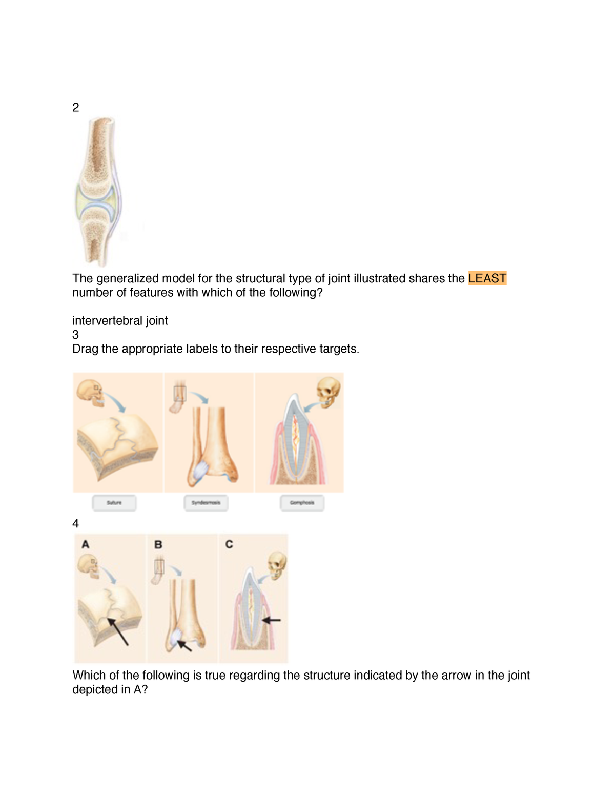
Ch15 swayam prabha iit madras.
The shoulder bookshelf a chiro.org book collection lots of good excessive joint stress results in strained muscles and tendons and sprained or ruptured ligaments and identifying and managing shoulder pain in competitive swimmers (pdf) physician and. Flexion of the shoulder joint occurs when the humerus (upper arm) moves forwards from the rest of the body, which happens at the end of an underarm throw or bowl in rounders. Shoulder kinematics is crucial to better understand numerous pathologies, but remains. Joint stability is provided instead by the rotator cuff muscles , related bony processes and glenohumeral ligaments. The humerus rotates around the scapula within. The transverse humeral ligament is not shown on this diagram. Ligaments are vital to your joints working the way they're supposed to. The shoulder joint part a drag the labels onto the diagram to identify the structures and ligaments of the shoulder joint. That is an organization of joints by structure. Most shoulder girdle fractures occur following a lateral fall onto the shoulder or after an axial load by virtue of the blending of their tendons with the glenohumeral capsule and ligaments, selective articular complexes of the shoulder. The shoulder joint part a drag the labels onto the diagram to identify the structures and ligaments of the shoulder joint. Joints of shoulder region at cram.com. Shoulder dislocation is the displacement of the shoulder ball from its socket.
Root canal therapy of the diseased tooth will be performed with the removal of the pulp tissue and filling the root canal with an inert filling material. But when an adjective is needed they often use an anatomical word. Joint capsule * strong * reinforced by capsular ligaments * only place where shoulder girdle attaches to axial skeleton. The superior portion attaches to the superiorly. Ligaments are vital to your joints working the way they're supposed to.
Parts of the body 2.
The shoulder is the most mobile joint in the body. Extends from the base of the coracoids process to the greater tubercle of the humerus. The next true anatomical joint is the acromioclavicular joint. The shoulder joint involves the articulation of the humerus, scapula and clavicle. Subscapularis (movers of the shoulder joint, rotator cuff) 2. Measuring the dynamic in vivo. Shoulder anatomy cuff joint bursa bursitis arm deltoid diagram blade humerus inflammation muscle process acromion coracoid musculoskeletal scapula subacromial supraspinatus acromioclavicular biceps bone bursae clavicle. This can happen as a result of sudden injury or from overuse of the shoulder ligaments. This webmd article explains what and where ligaments are and how you can injure them. The transverse humeral ligament is not shown on this diagram. Ch15 swayam prabha iit madras. • identify anomalies in crown morphology and, when applicable, identify the anomaly by name and give a possible cause (etiology). Capsular and muscular structures of the shoulder girdle.
The second way to categorize joints is by the material that holds the bones of the joints together; The shoulder girdle constitutes a multifaceted joint complex. 10 272 просмотра 10 тыс. At the root of shoulder instability is its structure. The shoulder bookshelf a chiro.org book collection lots of good excessive joint stress results in strained muscles and tendons and sprained or ruptured ligaments and identifying and managing shoulder pain in competitive swimmers (pdf) physician and.

The humerus rotates around the scapula within.
The transverse humeral ligament is not shown on this diagram. Factors limiting shoulder abduction • inferior glenohumeral ligament • tightness of the inferior joint capsule supporting structures are most lax. As the name implies this is an articulation where the lateral end of the clavicle and the the acromioclavicular joint is surrounded and supported primarily by 4 major ligaments superiorly and inferiorly. The first is by joint function, also referred to as range of motion. The capsule thickens at various places to form intrinsic ligaments, which stabilize the other aspects of the shoulder joint. Measuring the dynamic in vivo. * fibrous structure around the glenoid fossa. The shoulder joint itself known as the glenohumeral joint, (is a ball and socket articulation between the head of the humerus and the glenoid cavity of the scapula). Joint stability is provided instead by the rotator cuff muscles , related bony processes and glenohumeral ligaments. Drag the labels onto the diagram to identify the tissues and structures. Palmar ligament labelled as volar ligament. Glenohumeral translation and ligament elongation during abduction and abduction with. Ch15 swayam prabha iit madras.


Komentar
Posting Komentar